From the 2026 Summer Journal of the Colorado Dental Association, By Peter A. McIntyre, D.D.S.
Imagine two people who both stop breathing the same number of times each night. On paper, a sleep test gives them the same score, the apnea-hypopnea index (AHI). The AHI is the number of times per hour that breathing pauses or grows very shallow. For years, that one number was treated as the whole story. But it isn’t. The AHI counts how often breathing is interrupted but it does not measure how much harm is caused by each interruption. And when it comes to the heart, the harm is what matters.
Why Counting Falls Short
Every time breathing pauses, blood oxygen drops, then climbs back. Some drops are small and short while others are deep and long. A small dip is like dipping a toe in cold water and a deep drop is like jumping all the way in. The body feels them very differently, even though a counter labels each as “one event.”
Doctors now have a better measure: the hypoxic burden. Instead of counting pauses, it adds up how far the oxygen falls and how long it stays down throughout the night. The AHI tells you how often; the hypoxic burden tells you how much. Large studies show it is the “how much” that lines up with strain on the heart over time.
A Picture Is Easier Than a Number
The drawing below shows the same idea two ways. The red area is the oxygen the body loses while sleeping; the larger the area, the more the heart works. Two people can have the same AHI and still have very different amounts of red.
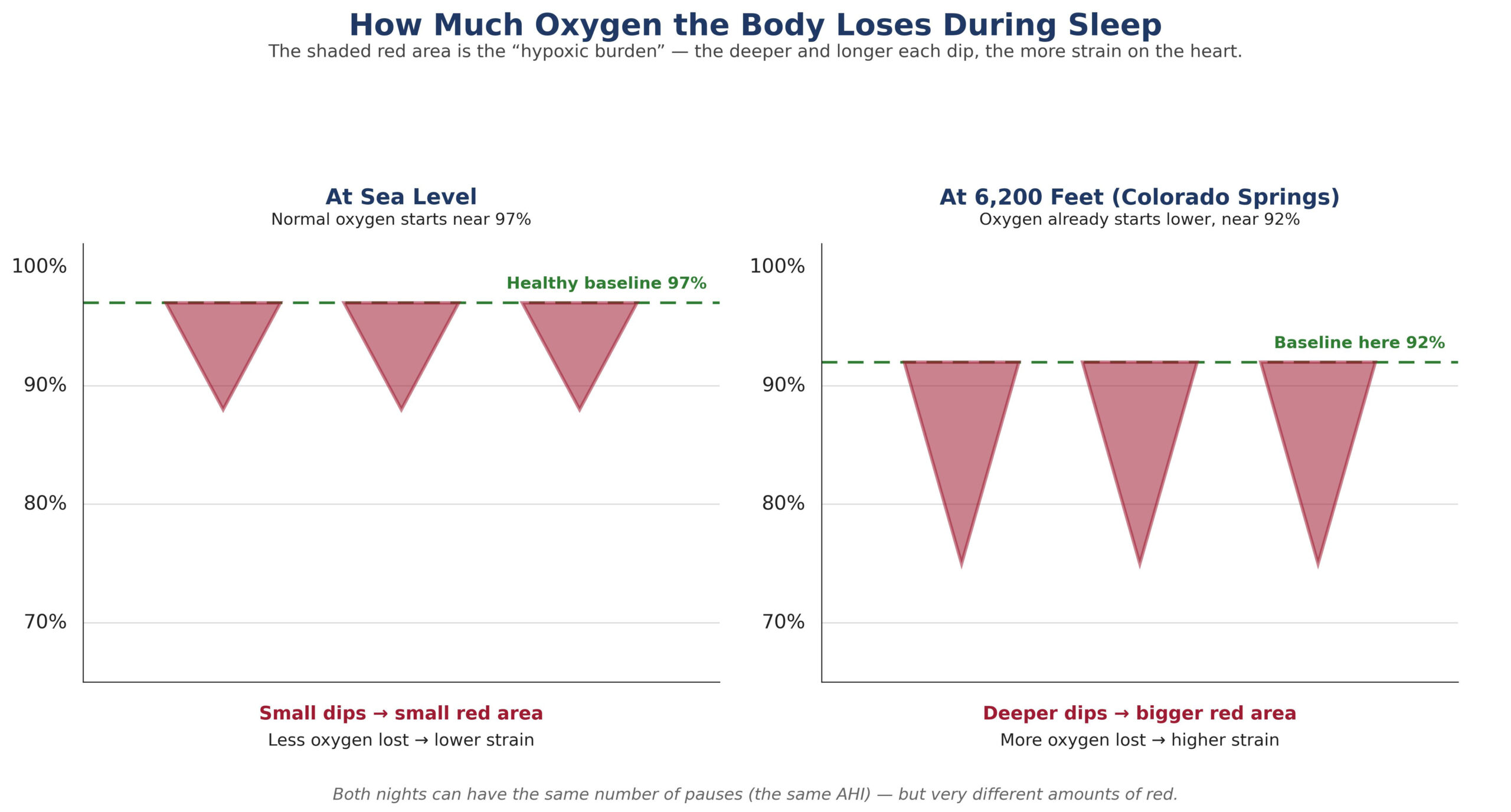
Figure 1. The same number of breathing pauses can cause very different amounts of oxygen loss.
This is not just theory. In our own reviewed cases, we have seen two people with nearly the same AHI whose hypoxic burdens told very different stories. One modest, one heavy – and it was the heavier burden, not the matching event count, that flagged the patient who needed closer attention. Reviewing cases this way, from the study through follow-up, is exactly what the metric is for.
Cardiopulmonary Coupling Study Readout
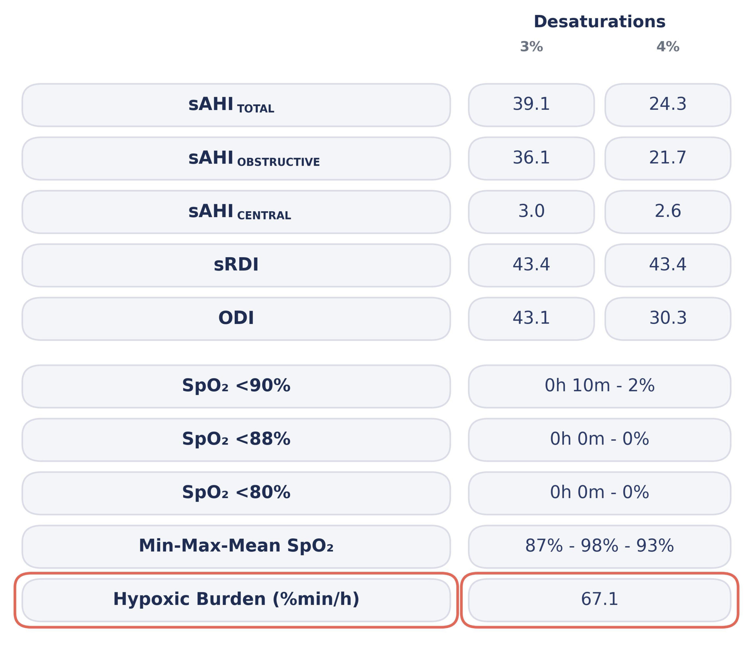
Because it reflects the severity of intermittent hypoxia rather than event frequency alone, hypoxic burden has been associated with cardiovascular risk in a manner that the AHI and percentage of time below 90% (T90) don’t consistently predict.
Why Living in Colorado Springs Changes the Picture
At sea level, a healthy sleeper’s oxygen sits around 97%. Colorado Springs sits at about 6,200 feet, where the air holds less oxygen, so a normal person here may start the night closer to 92%. Beginning lower, the dips land deeper, carving out a larger red area than at sea level. This also explains why T90 is misleading; at high altitude, a normal sleeper may cross it from thin air alone. The hypoxic burden avoids this trap because it measures each person against their own starting point.
Who Does What
Roles matter. The sleep physician diagnoses apnea and, with the help of the dentist, decides treatment. The dentist’s job is supportive: to recognize the signs, help the patient reach the right physician and read the sleep study sensibly when it returns. A dentist who understands the hypoxic burden, and does not stop at the AHI, helps the whole team see the patient’s real risk.
The Bottom Line
Do not let a single number tell the whole story. Counting the pauses is not the same as measuring the harm. Ask the better question — not , “How many times did breathing stop?” but “How much oxygen did the body lose, and is this heart protected?” That is what the hypoxic burden was built to answer.
About the Author
Peter A. McIntyre, D.D.S., owns Briargate Advanced Family Dental in Colorado Springs and serves on the faculty of the Pikes Peak State College Dental Hygiene Program. He has a background in respiratory therapy and holds preceptorship training in the Bale/Doneen Method for cardiovascular risk reduction, integrating cardiopulmonary coupling sleep analysis and CBCT-based airway assessment into his clinical workflow. He is a member of the Advanced Airway Alliance, a group of clinicians who continually share and monitor sleep cases together traversing the data through planning and treatment.
Further Reading
Azarbarzin A, Sands SA, Stone KL, et al. The hypoxic burden of sleep apnoea predicts cardiovascular disease-related mortality: the Osteoporotic Fractures in Men Study and the Sleep Heart Health Study. Eur Heart J. 2019;40(14):1149–1157.
McEvoy RD, Antic NA, Heeley E, et al. CPAP for prevention of cardiovascular events in obstructive sleep apnea (SAVE). N Engl J Med. 2016;375(10):919–931.
Sánchez-de-la-Torre M, Sánchez-de-la-Torre A, Bertran S, et al. Effect of obstructive sleep apnoea and its treatment on cardiovascular events after acute coronary syndrome (ISAACC): a randomized controlled trial. Lancet Respir Med. 2020;8(4):359–367.
Peker Y, Glantz H, Eulenburg C, et al. Effect of positive airway pressure on cardiovascular outcomes in coronary artery disease patients with nonsleepy obstructive sleep apnea: the RICCADSA randomized controlled trial. Am J Respir Crit Care Med. 2016;194(5):613–620.
Punjabi NM. The epidemiology of adult obstructive sleep apnea. Proc Am Thorac Soc. 2008;5(2):136–143.
Glossary of Sleep Studies and Terms
Polysomnography (PSG). An overnight, attended sleep study performed in a laboratory that records brain waves, breathing, oxygen, heart rhythm and muscle activity. It is the most complete test and the reference standard for diagnosing sleep-disordered breathing.
Home Sleep Apnea Test (HSAT). A simplified study the patient wears at home, recording airflow, breathing effort, oxygen and heart rate. Convenient and lower in cost, though it records fewer signals than full PSG.
Cardiopulmonary Coupling (CPC). An analysis derived from the heart rate and breathing signals that maps the coordination between the heart and lungs during sleep. It is sensitive to the autonomic (sympathetic) disruption that event-counting can miss.
Apnea-Hypopnea Index (AHI). The number of breathing pauses (apneas) and shallow-breathing events (hypopneas) per hour of sleep. It counts how often breathing is disturbed but does not measure how much oxygen is lost.
Hypoxic Burden. A measure of the total oxygen lost during sleep — the depth of each desaturation multiplied by its duration, summed across the night. It reflects how much, not just how often, and tracks more closely with cardiovascular risk.
Oxygen Desaturation Index (ODI 3%). The number of times per hour that blood oxygen falls at least 3% below the patient’s own baseline. Because it is anchored to each individual, it stays reliable at altitude.
T90. The percentage of sleep time spent with oxygen below 90%. Because 90% is a fixed line, T90 can mislead at higher elevations where baseline oxygen is already lower.